New Patients
(732) 813-3564
Existing Patients
(732) 639-3495

At the office of Elegant Dental Arts in Freehold, NJ, we prioritize precise diagnosis and thoughtful planning. Cone-beam computed tomography (CBCT) is one of the diagnostic tools that helps our team see the full picture — literally — by producing three-dimensional images of the teeth, jaws, and surrounding anatomy. When combined with a thorough clinical exam, CBCT delivers actionable information that supports safer, more predictable care for patients across many treatment types.
CBCT is not intended to replace routine 2D radiographs or a clinician’s judgment, but it adds an important layer of detail when anatomy, pathology, or surgical planning demand a higher level of visualization. The technology offers a clear advantage in complex cases, and when used appropriately it enhances diagnostic confidence while preserving patient comfort and safety.
Conventional dental X-rays are excellent for many routine needs, but they show structures in two dimensions. CBCT overcomes overlap and distortion by capturing volumetric data that can be examined in axial, coronal, and sagittal planes. This 3D perspective clarifies relationships between roots, nerve canals, sinus cavities, and bony contours — information that is often essential when a single plane view leaves uncertainty.
CBCT can reveal subtle anatomic variations such as accessory canals, complex root curvatures, or thin cortical plates that are difficult to appreciate on 2D films. It also helps detect and localize lesions or anatomical changes that might otherwise be missed until they become more advanced. For clinicians, this means better-informed decisions; for patients, it means conditions can be identified earlier and managed more precisely.
Importantly, CBCT provides true spatial measurements, which are critical when precise distances and angulations guide treatment. These quantifiable data are used for planning and risk assessment, reducing guesswork and improving procedural predictability while complementing the clinical examination.
When planning surgical procedures — particularly implant placement — CBCT enables the clinician to virtually evaluate bone volume, density, and the position of vital structures like the inferior alveolar nerve and maxillary sinus. This preoperative insight allows for more accurate implant sizing and positioning, and it supports the design of surgical guides when indicated.
In endodontics, CBCT assists in identifying complex canal systems, root fractures, and periapical pathology that may not be evident on traditional films. For oral and maxillofacial surgery, it informs extractions, bone graft planning, and assessment of cysts or other pathologic conditions. Orthodontic and airway assessments can also benefit from 3D data when evaluating skeletal relationships and airway volume.
By integrating CBCT data with digital planning tools and intraoral scans, dental teams can create coordinated, multidisciplinary treatment plans. This integration supports clearer communication among providers and with patients, helping everyone understand the rationale behind recommended care and the expected steps of treatment.
CBCT examinations are designed to be efficient and patient-friendly. Scans typically take only a matter of seconds to capture, and modern units offer adjustable fields of view so clinicians can limit exposure to the region of interest. Focusing on a specific area — rather than imaging the entire head — reduces radiation dose while providing the high-resolution detail needed for diagnosis.
Patient positioning is straightforward, and the short scan time minimizes movement artifacts that could compromise image quality. Our team follows established safety protocols and adheres to the ALARA principle (as low as reasonably achievable) to ensure every exam is justified and optimized for diagnostic value.
Because CBCT units used in dental settings are different from full medical CT scanners, they typically deliver lower doses of radiation for targeted dental applications. The choice to image with CBCT is made carefully, with consideration of clinical necessity, alternatives, and the potential benefit to the patient’s treatment outcome.
CBCT produces a wealth of information, but like all diagnostic tools it must be interpreted within a clinical context. Recognizing normal variations, incidental findings, and artifacts requires training and experience. Our clinicians review 3D datasets alongside a patient’s history and examination findings to draw clinically relevant conclusions and avoid overinterpretation of incidental details.
Specialized software allows clinicians to manipulate views, take measurements, and generate cross-sectional slices that clarify complex anatomy. These capabilities are valuable, but they demand skill to translate images into safe, practical treatment plans. When needed, collaboration with radiologists or specialists is part of a prudent diagnostic process to ensure comprehensive evaluation.
Equally important is communication: clear explanations of what the images show and how that information affects treatment choices help patients make informed decisions. We focus on presenting findings in plain language, outlining the clinical implications, and answering questions so patients understand their condition and the proposed care pathway.
CBCT has become a cornerstone of modern, digitally driven dental care. When combined with intraoral scanning and CAD/CAM technologies, 3D imaging supports streamlined workflows for restorative, surgical, and prosthetic treatments. This digital alignment can shorten treatment timelines and increase the accuracy of laboratory-fabricated restorations and surgical guides.
For multidisciplinary cases, CBCT bridges specialties by providing a consistent reference dataset that surgeons, restorative dentists, and lab technicians can use to coordinate. Whether planning an implant-supported restoration, evaluating a potential surgical approach, or assessing complex anatomy for endodontic retreatment, the shared visual information enhances team collaboration.
Finally, CBCT aids in monitoring healing and assessing treatment outcomes when repeat imaging is clinically indicated. Used judiciously, it offers a powerful means to document changes over time and to confirm that treatment objectives have been met without relying solely on two-dimensional images.
In summary, CBCT is a sophisticated diagnostic tool that supports more precise, informed dental care across a variety of clinical situations. When combined with experienced interpretation and a comprehensive clinical exam, three-dimensional imaging enhances planning, communication, and outcomes. For patients seeking clarity about their diagnosis or treatment options, please contact us for more information.
Cone-beam computed tomography, commonly called CBCT, is a three-dimensional imaging technology that captures volumetric data of the teeth, jaws, and surrounding structures. Unlike traditional two-dimensional films, CBCT produces cross-sectional views that let clinicians evaluate anatomy in axial, coronal, and sagittal planes. This 3D perspective clarifies spatial relationships and provides true measurements that support precise diagnosis and planning.
In dental practice, CBCT is used across many specialties including implant planning, endodontics, oral surgery, orthodontics, and airway assessment. The information gleaned from a CBCT scan helps clinicians identify anatomic variations, localize pathology, and assess bone volume and density before treatment. At Elegant Dental Arts in Freehold, NJ, CBCT is integrated with clinical exams to improve diagnostic confidence and treatment predictability.
Standard dental X-rays produce two-dimensional images that are excellent for many routine needs but can suffer from overlap and distortion of structures. CBCT overcomes these limitations by acquiring a volumetric dataset that can be reformatted into multiple planar and three-dimensional views. This removes superimposition and reveals depth, angulation, and the true course of roots, canals, and nerve channels.
The ability to take precise measurements and inspect anatomy from different angles is a key advantage of CBCT, especially when a single-plane view leaves uncertainty. However, CBCT complements rather than replaces conventional radiographs and clinical judgment, since 2D films remain useful for many diagnostic and monitoring tasks. Clinicians choose the imaging modality that best balances information needs and patient exposure.
A dentist may recommend CBCT when the clinical situation requires detailed three-dimensional information that cannot be obtained from routine radiographs. Common indications include complex implant planning, assessment of impacted or difficult extractions, evaluation of suspected root fractures, and investigation of unusual pathology or anatomy. CBCT is also useful for airway analysis and orthodontic cases where skeletal relationships need precise assessment.
The decision to use CBCT is case-specific and follows the ALARA principle, meaning the exam is justified only when the expected diagnostic benefit outweighs the exposure. Your clinician will consider alternatives, the field of view required, and whether focused imaging of a region of interest can provide the needed detail. When appropriate, CBCT leads to safer, more predictable treatment planning.
CBCT systems used in dental settings are designed to deliver a focused beam and typically expose patients to lower doses than conventional medical CT scanners for comparable areas. Modern units offer adjustable fields of view and optimized protocols to limit radiation to the region of interest, which helps minimize overall exposure. Clinicians follow established safety guidelines and apply the ALARA concept to ensure each scan is justified and optimized.
Radiation dose varies with factors such as field of view, image resolution, and machine settings, so discussing these details with your provider is reasonable when a scan is recommended. For most dental applications, the diagnostic benefit of CBCT—such as avoiding surgical complications or detecting pathology early—outweighs the small incremental exposure. If you have specific concerns about radiation, your dentist can explain the rationale and any alternatives available.
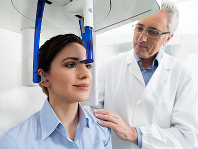
A CBCT appointment is typically quick and straightforward, with most commercial dental scans captured in a matter of seconds. You will be positioned in the unit—either seated, standing, or in some models lying down—and asked to remain still while the scanner rotates around your head to collect the images. Patient comfort is usually high because the procedure is noninvasive and does not require contrast agents or special preparation.
To optimize image quality you may be asked to remove metal objects such as jewelry or removable dental appliances from the area of interest. The team will tailor the field of view and resolution to the diagnostic need, which helps reduce exposure and focus on the relevant anatomy. After the scan, the dataset is available immediately for review and planning.
Interpreting CBCT datasets requires training and experience to recognize normal anatomic variation, artifacts, and clinically relevant findings. In many dental practices the treating clinician reviews the images alongside the patient's history and examination to draw practical conclusions for treatment. For complex or ambiguous findings, collaboration with oral and maxillofacial radiologists or specialists can provide an added level of diagnostic certainty.
Once interpreted, CBCT data are used to guide specific treatment steps such as implant placement, endodontic retreatment, or surgical planning. The images also support patient education by visually demonstrating anatomy and explaining proposed interventions. Clear communication about what the scan shows and how it affects options is an important part of responsible care.
CBCT provides critical information for implant planning by revealing bone volume, density, and the spatial relationship of vital structures like the inferior alveolar nerve and maxillary sinus. This allows clinicians to select appropriate implant length, diameter, and angulation while avoiding anatomical hazards. Accurate measurements from CBCT reduce guesswork and help determine whether bone grafting or staged approaches are necessary.
CBCT datasets can also be integrated with digital impression data to design surgical guides and prosthetic components that fit precisely. The combination of 3D imaging and digital workflows increases predictability, shortens chair time in some cases, and facilitates communication with laboratory technicians and specialists. Overall, CBCT supports safer and more efficient implant care when used judiciously.
Yes, CBCT can reveal conditions that are difficult to appreciate on two-dimensional films, such as accessory root canals, small periapical lesions obscured by anatomical overlap, and subtle root fractures. The absence of superimposed structures in a 3D dataset makes it easier to localize pathology and evaluate bony contours or cortical breaches. This enhanced visibility can lead to earlier detection and more targeted treatment.
However, not every concern requires CBCT, and incidental findings that are clinically insignificant can appear on a volumetric scan. Experienced interpretation helps distinguish meaningful pathology from benign variants or imaging artifacts, and when appropriate additional consultation with a radiologist or specialist ensures comprehensive evaluation. CBCT is a powerful tool but must be applied and interpreted thoughtfully.
CBCT is subject to limitations such as lower soft-tissue contrast compared with medical CT and susceptibility to artifacts from metal restorations or patient movement. Beam hardening and scatter around metal objects can create streaks or distortions that obscure adjacent structures, and small field-of-view scans may miss pathology outside the imaged region. Recognizing these constraints is important to avoid overreliance on a single dataset.
To mitigate artifacts clinicians select appropriate imaging parameters, remove removable metal when feasible, and use stabilization techniques to limit motion. When CBCT is insufficient for a specific diagnostic question, referral for alternative imaging or radiologic consultation may be warranted. Sound clinical judgment determines when CBCT will add meaningful value to patient care.
CBCT functions as a central reference in many digital workflows by providing a consistent 3D dataset that can be merged with intraoral scans, facial scans, and CAD/CAM designs. This interoperability allows restorative dentists, surgeons, and laboratories to coordinate implant position, prosthetic contours, and surgical guides with greater precision. Shared visual information streamlines planning meetings and helps align expectations across the treatment team.
For patients, the integrated approach clarifies treatment steps and anticipated outcomes and can shorten timelines when components are digitally designed and pre-fabricated. If you have questions about how 3D imaging will be used in your case, contact Elegant Dental Arts in Freehold, NJ to discuss how CBCT may support a coordinated plan tailored to your needs.